This scanning electron micrograph (SEM) depicted a number of red
By A Mystery Man Writer
Description
Download this stock image: This scanning electron micrograph (SEM) depicted a number of red blood cells found enmeshed in a fibrinous matrix on the luminal surface of an indwelling vascular catheter; Magnified 11432x Note the biconcave cytomorphologic shape of each erythrocyte, which increases the surface area of these hemoglobin-filled cells, thereby, promoting a greater degree of gas exchange, which is their primary function in an in vivo setting. In their adult phase, these cells possess no nucleus. What appears to be irregularly-shaped chunks of debris, are actually fibrin clumps, which when inside the living organi - 2BE0H0B from Alamy's library of millions of high resolution stock photos, illustrations and vectors.
![This scanning electron micrograph (SEM) depicted a number of red](https://ars.els-cdn.com/content/image/3-s2.0-B9780123847300002159-f00215-01-9780123847300.jpg)
Scanning Electron Microscope - an overview
![This scanning electron micrograph (SEM) depicted a number of red](https://upload.wikimedia.org/wikipedia/commons/thumb/d/d7/DDC-SEM_of_calcified_particle_in_cardiac_tissue_-_BW_-_2.jpg/220px-DDC-SEM_of_calcified_particle_in_cardiac_tissue_-_BW_-_2.jpg)
Scanning electron microscope - Wikipedia
![This scanning electron micrograph (SEM) depicted a number of red](https://upload.wikimedia.org/wikipedia/commons/thumb/c/cb/Surface_of_a_kidney_stone.jpg/196px-Surface_of_a_kidney_stone.jpg)
Scanning electron microscope - Wikipedia
![This scanning electron micrograph (SEM) depicted a number of red](https://render.fineartamerica.com/images/rendered/default/duvet-cover/images-medium/40-red-blood-cells-sem-science-source.jpg?&targetx=259&targety=311&imagewidth=325&imageheight=221&modelwidth=844&modelheight=844&backgroundcolor=242424&orientation=0&producttype=duvetcover-queen)
Red Blood Cells, Sem #40 Duvet Cover by Science Source - Science
![This scanning electron micrograph (SEM) depicted a number of red](https://t1.thpservices.com/previewimage/gallil/e911aaa5d2938a270e28a325c83daf23/sjb-4384-206.jpg)
This scanning electron micrograph SEM revealed some of the
![This scanning electron micrograph (SEM) depicted a number of red](https://upload.wikimedia.org/wikipedia/commons/a/a4/Misc_pollen.jpg)
Scanning electron microscope - Wikipedia
![This scanning electron micrograph (SEM) depicted a number of red](https://www.superstock.com/cdn/4128/Comp/4128R-30461.webp)
Activated human macrophage, coloured scanning electron micrograph (SEM). Magnification: x2,700 when printed at 10 centimetres wide. - SuperStock
![This scanning electron micrograph (SEM) depicted a number of red](https://t1.thpservices.com/previewimage/gallil/8f7028dd8798cb681e81820f16651eec/sjb-4384-108.jpg)
This scanning electron micrograph SEM revealed some of the
![This scanning electron micrograph (SEM) depicted a number of red](https://media.gettyimages.com/id/151052558/photo/the-last-step-of-blood-coagulation-is-marked-by-the-transformation-of-a-soluble-blood-protein.jpg?s=612x612&w=gi&k=20&c=7zcVAfmbwISZiodCbDFIPKk-QHhuQwcaLB9iqutne7s=)
106 Blood Clot Fibrin Stock Photos, High-Res Pictures, and Images
![This scanning electron micrograph (SEM) depicted a number of red](https://render.fineartamerica.com/images/rendered/small/flat/sticker/images-medium/3-red-blood-cells-and-acanthocyte-sem-science-source.jpg?transparent=0&targetx=0&targety=160&imagewidth=1000&imageheight=679&modelwidth=1000&modelheight=1000&backgroundcolor=1C1C1C&orientation=0&producttype=sticker-3-3&brightness=84&stickerbackgroundcolor=transparent)
Red Blood Cells And Acanthocyte, Sem #3 Photograph by Science
![This scanning electron micrograph (SEM) depicted a number of red](https://l450v.alamy.com/450v/2be0h07/this-highly-enlarged-scanning-electron-micrograph-sem-depicted-a-closer-look-at-the-details-exhibited-by-of-number-of-red-blood-cells-found-enmeshed-in-a-fibrinous-matrix-on-the-luminal-surface-of-an-indwelling-vascular-magnified-11397x-in-this-instance-the-indwelling-catheter-was-a-tube-that-was-left-in-place-creating-a-patent-portal-directly-into-a-blood-vessel-note-the-biconcave-cytomorphologic-shape-of-each-erythrocyte-which-increases-the-surface-area-of-these-hemoglobin-filled-cells-thereby-promoting-a-greater-degree-of-gas-exchange-which-is-their-primary-function-in-an-in-vivo-2be0h07.jpg)
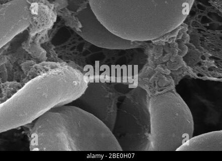
This highly enlarged scanning electron micrograph (SEM) depicted a closer look at the details exhibited by of number of red blood cells found enmeshed in a fibrinous matrix on the luminal surface of an indwelling vascular; Magnified 11397x. In this instance
![This scanning electron micrograph (SEM) depicted a number of red](https://static.cambridge.org/content/id/urn%3Acambridge.org%3Aid%3Aarticle%3AS1431927620001713/resource/name/S1431927620001713_figAb.png?pub-status=live)
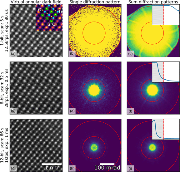
Fast Pixelated Detectors in Scanning Transmission Electron Microscopy. Part I: Data Acquisition, Live Processing, and Storage, Microscopy and Microanalysis
![This scanning electron micrograph (SEM) depicted a number of red](https://t1.thpservices.com/previewimage/gallil/479564f0140df62771e1603af72fa0e7/sjb-4384-126.jpg)
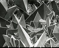
ACANTHOCYTE, RED BLOOD CELL
This scanning electron micrograph
![This scanning electron micrograph (SEM) depicted a number of red](https://c8.alamy.com/comp/E7TW0P/orbulina-coloured-scanning-electron-micrograph-sem-of-the-shell-of-E7TW0P.jpg)
Electron micrograph hi-res stock photography and images - Alamy
![This scanning electron micrograph (SEM) depicted a number of red](https://www.researchgate.net/publication/263710781/figure/fig11/AS:669648890642449@1536668145914/Extracellular-matrix-changes-in-Flk1-cKO-mutants-A-D-Scanning-electron-microscopy-of.png)
Extracellular matrix changes in Flk1 cKO mutants. (A,D) Scanning
from
per adult (price varies by group size)