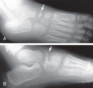
Foot X-ray of a 10 year-old male patient (white arrow indicates
By A Mystery Man Writer
Description
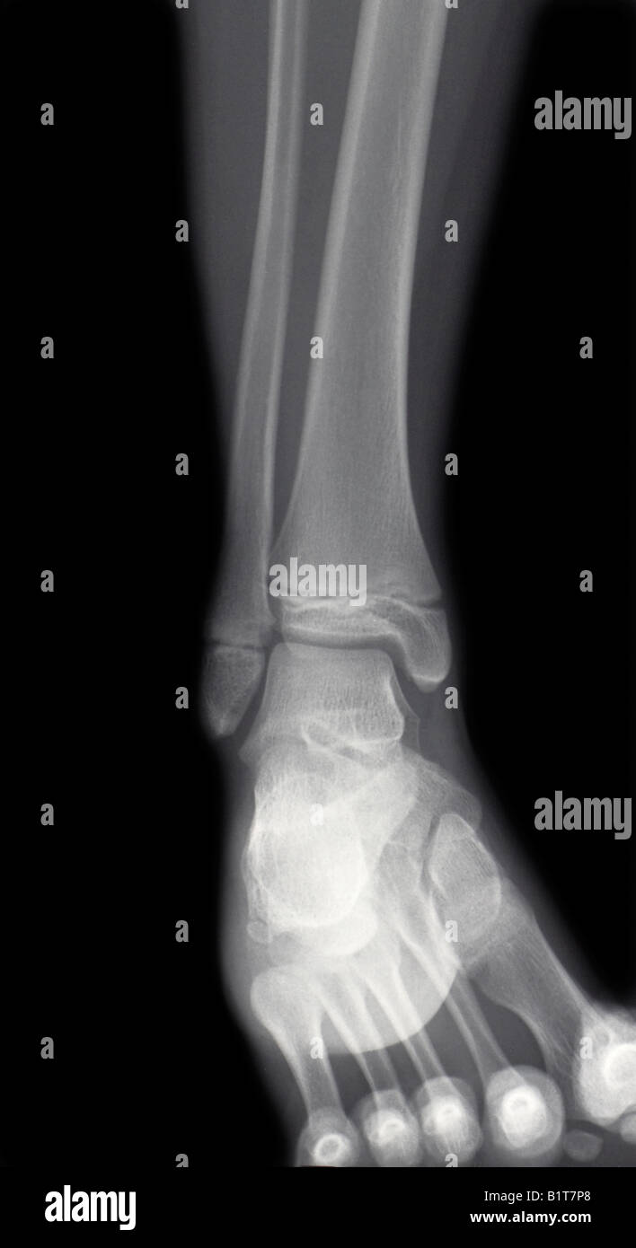
X ray of foot hi-res stock photography and images - Alamy

Ozlem Bilir's research works Recep Tayyip Erdoğan Üniversitesi, Rize and other places
Skeletal System
Gluteus Minimus Anatomy and Tear Patterns

Cervical adjacent segment disease: Risks and complications following cervical fusion – Caring Medical Florida

PDF) Sprain Injury in a Child: Where is the Fracture Line?

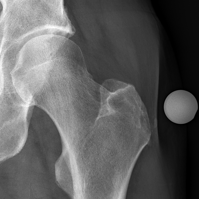
X-ray of the right foot (case no. 1) showing a radiolucent lesion of

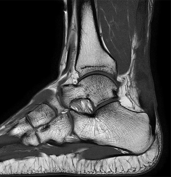
Radiographic analysis of adult ankle fractures using combined Danis-Weber and Lauge-Hansen classification systems
/wp-content/uploads/2017/01/1a-1.jpg
from
per adult (price varies by group size)