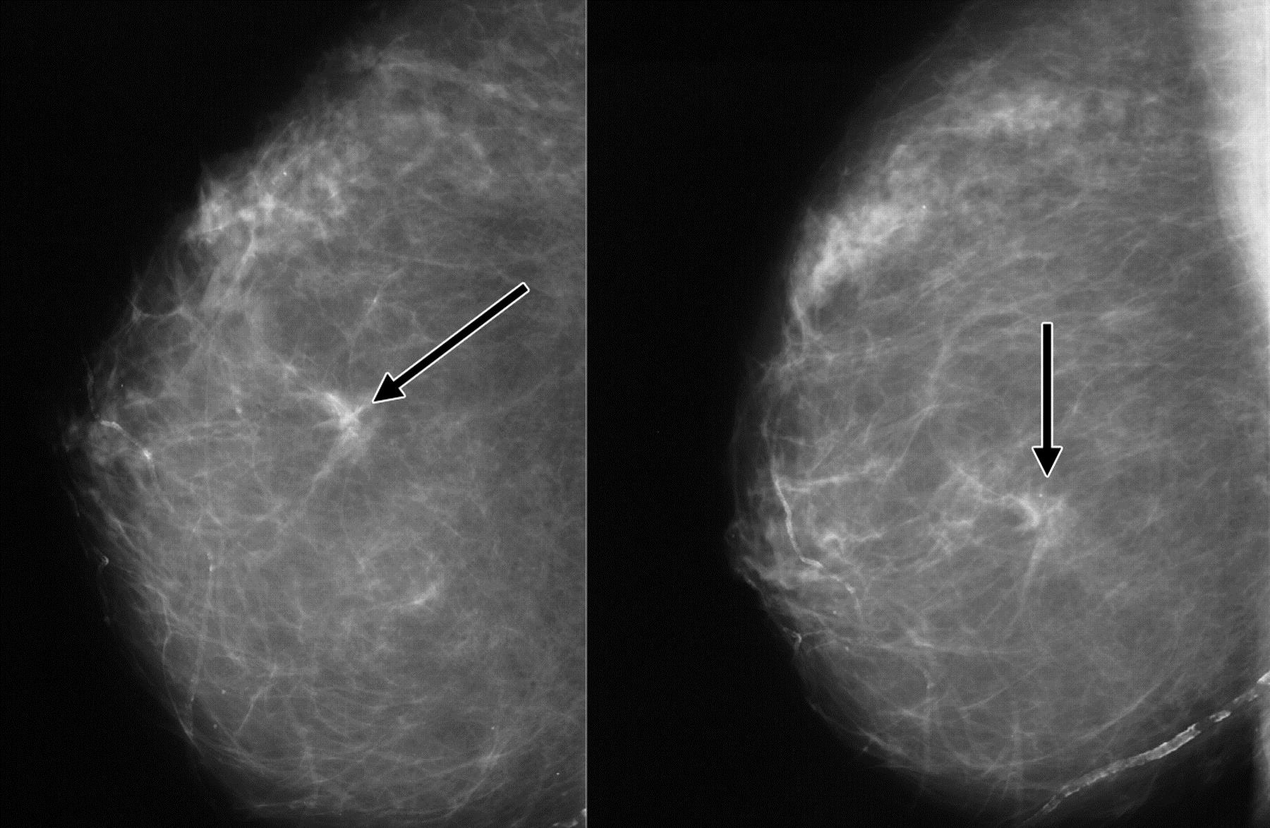
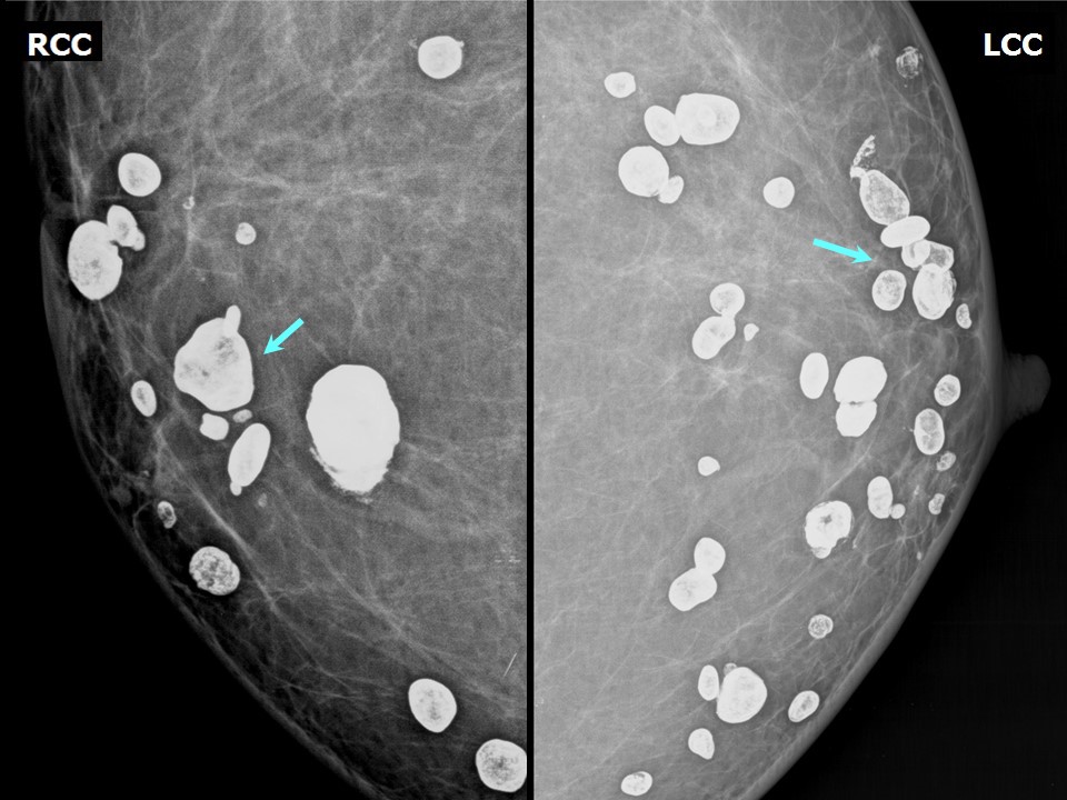
Calcification and mass abnormalities in breast mammogram scans
By A Mystery Man Writer
Description
Download scientific diagram | Calcification and mass abnormalities in breast mammogram scans. The calcification distribution depicts tiny flecks of calcium as small white regions on the left side, while the mass is shown as a smooth, well-defined border on the right side. from publication: Multi-Graph Convolutional Neural Network for Breast Cancer Multi-Task Classification | Mammography is a popular diagnostic imaging procedure for detecting breast cancer at an early stage. Various deep learning (DL) approaches to breast cancer detection incur high costs and are prone to classify incorrectly. Therefore, they are not sufficiently reliable to | Breast Cancer, Convolution and Classification | ResearchGate, the professional network for scientists.
Atlas of breast cancer early detection
Atlas of breast cancer early detection

Mohamed IBRAHIM, Master of Science
Comparison between Semi-Supervised GrowCut segmentation and ground
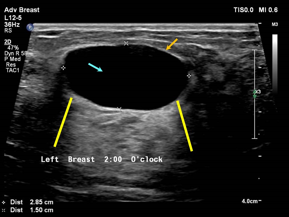
Breast Cancer Ultrasonography: Practice Essentials, Role of Ultrasonography in Screening, Breast Imaging Reporting and Data System
:max_bytes(150000):strip_icc()/lateral-mammogram-of-female-breast-with-tumor-92263689-813095ee469b45eabfc9f5f4747758ed.jpg)
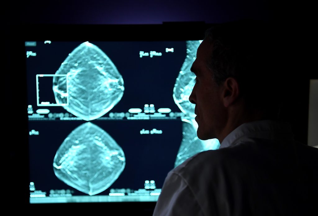
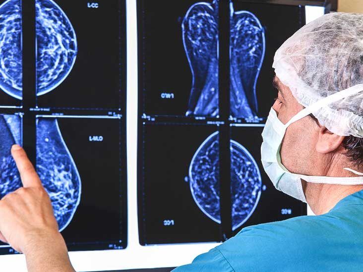
Mammogram Images: Normal and Abnormal

Brendan JENNINGS, Head of Graduate Studies
Breast Cancer Signs, Symptoms and Understanding an Imaging Report

Breast imaging-reporting and data system (BI-RADS), Radiology Reference Article

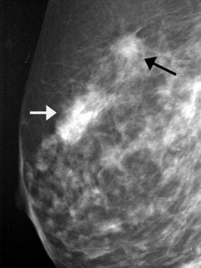
SA features on mammography. (A) Mammography depicts an 11 mm  12 mm

Breast arterial calcification on mammography and risk of coronary artery disease: a SCOT-HEART sub-study - ScienceDirect

Brendan JENNINGS, Head of Graduate Studies
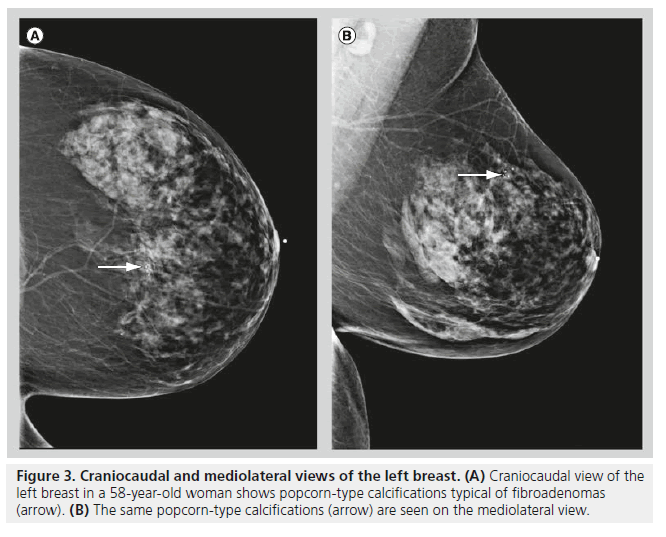
Mammography of breast calcifications
Understanding Your Mammogram Results

Calcification and mass abnormalities in breast mammogram scans
from
per adult (price varies by group size)