Ultrasound imaging
By A Mystery Man Writer
Description
Ultrasound imaging - Download as a PDF or view online for free
Ultrasound uses high frequency sound waves to visualize internal structures. It works by transmitting sound waves into the body using a transducer probe, which detects the echoes as they bounce off tissues and organs. The echoes are processed to form images on the ultrasound machine screen in real-time. Common applications include obstetrics, cardiology, and urology. The Philips HD11 is an ultrasound system with curvilinear, linear, and phased array probes for different exams. It provides grey scale, Doppler, and color imaging modes. Ultrasound has benefits of being non-invasive, portable, and having no radiation, but has limitations of being operator dependent and unable to penetrate bone.
Ultrasound uses high frequency sound waves to visualize internal structures. It works by transmitting sound waves into the body using a transducer probe, which detects the echoes as they bounce off tissues and organs. The echoes are processed to form images on the ultrasound machine screen in real-time. Common applications include obstetrics, cardiology, and urology. The Philips HD11 is an ultrasound system with curvilinear, linear, and phased array probes for different exams. It provides grey scale, Doppler, and color imaging modes. Ultrasound has benefits of being non-invasive, portable, and having no radiation, but has limitations of being operator dependent and unable to penetrate bone.

Enhancing Safety with Ultrasound Imaging — Dr. Heather Friedman ND, LAc

Fetal Pictures of Ultrasounds Gallery

Cincinnati Children's Excels in Ultrasound Imaging - Radiating Hope

Ultrasound Imaging: What Is Inside? - Online Course - Futurelearn
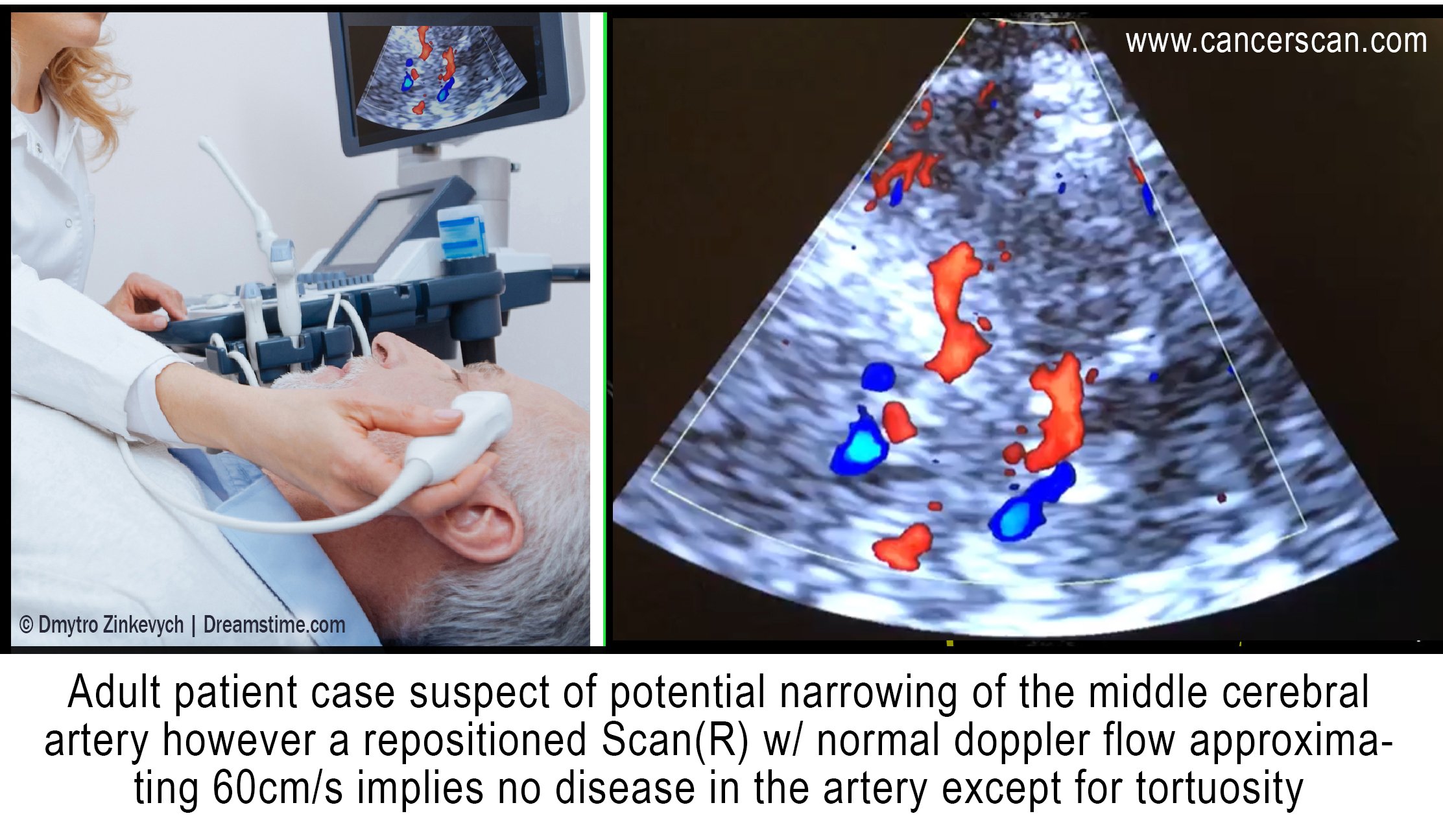
2021 Review: Transcranial Doppler Ultrasound Imaging

Through-needle all-optical ultrasound imaging in vivo: a preclinical swine study
DVD is designed to demonstrate musculoskeletal pathologies that require joint movement, positioning, or muscle contraction for accurate diagnosis.
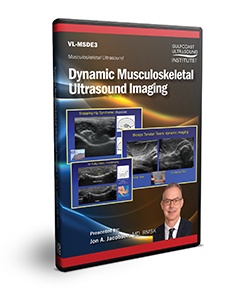
Dynamic Musculoskeletal Ultrasound Imaging
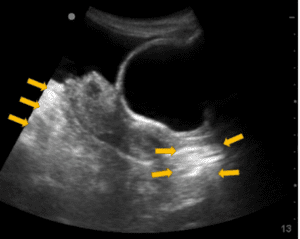
An ultrasound imaging artifact, Case Studies

The Science in Ultrasound Imaging And its Importance In OB-GYN
from
per adult (price varies by group size)
.jpg)