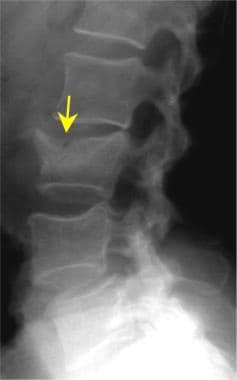
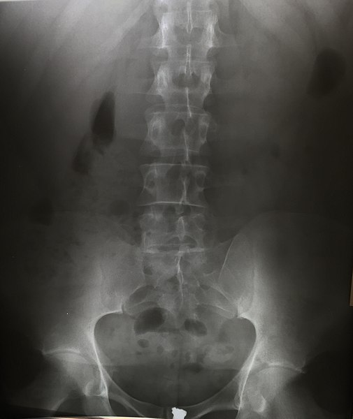
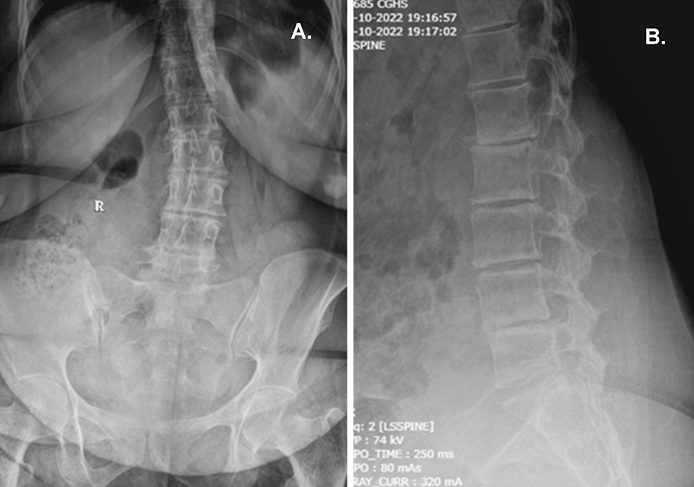
Standing anteroposterior and lateral X-rays of the dorso-lumbar spine
By A Mystery Man Writer
Description
Download scientific diagram | Standing anteroposterior and lateral X-rays of the dorso-lumbar spine showing a failure of the pedicular screws at T11. Note the iatrogenic flat-back deformity with loss of sagittal spine alignment and +ve sagittal vertical axis. from publication: Acute Paraplegia Secondary to Thoracic Disc Herniation of the Adjacent Segment Following Thoracolumbar Fusion and Instrumentation | Proximal junctional disease is a well-recognized postoperative phenomenon in adults who are undergoing long thoracolumbar fusion and instrumentation, and is attributed to increased a junctional stress concentration. In general, the onset of symptoms in these patients is | Paraplegia, Fusion and Segmentation | ResearchGate, the professional network for scientists.

Standing AP and lateral lumbar spine radiographs demonstrating
Xray cervical spine AP and lateral shows straightening of cervical spine? What does it mean? - Quora

Standing anteroposterior (A) and lateral (B) radiographs of the

Technique of Dorso-Lumber & Lumbo-Dorsal Spine (Ep 65)

Ramzi MOUCHARAFIEH, Professor (Full)
Lumbar Spine Trauma Imaging: Practice Essentials, Radiography, Computed Tomography
Approach to Thoracic and Lumbar Spine X-ray

Lumbar-pelvic-femoral balance on sitting and standing lateral radiographs - ScienceDirect
Cureus, Alkaptonuria Presenting With Lumbar Disc Herniation: A Case Report
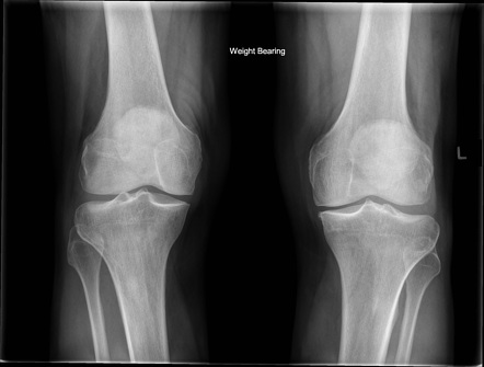
Knee (AP weight-bearing view), Radiology Reference Article
from
per adult (price varies by group size)