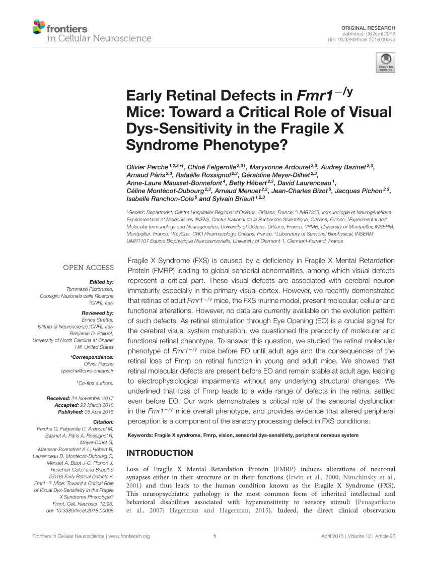
Optical Coherence Tomography: Imaging Mouse Retinal Ganglion Cells In Vivo
By A Mystery Man Writer
Description
Scientific Article | Structural changes in the retina are common manifestations of ophthalmic diseases.
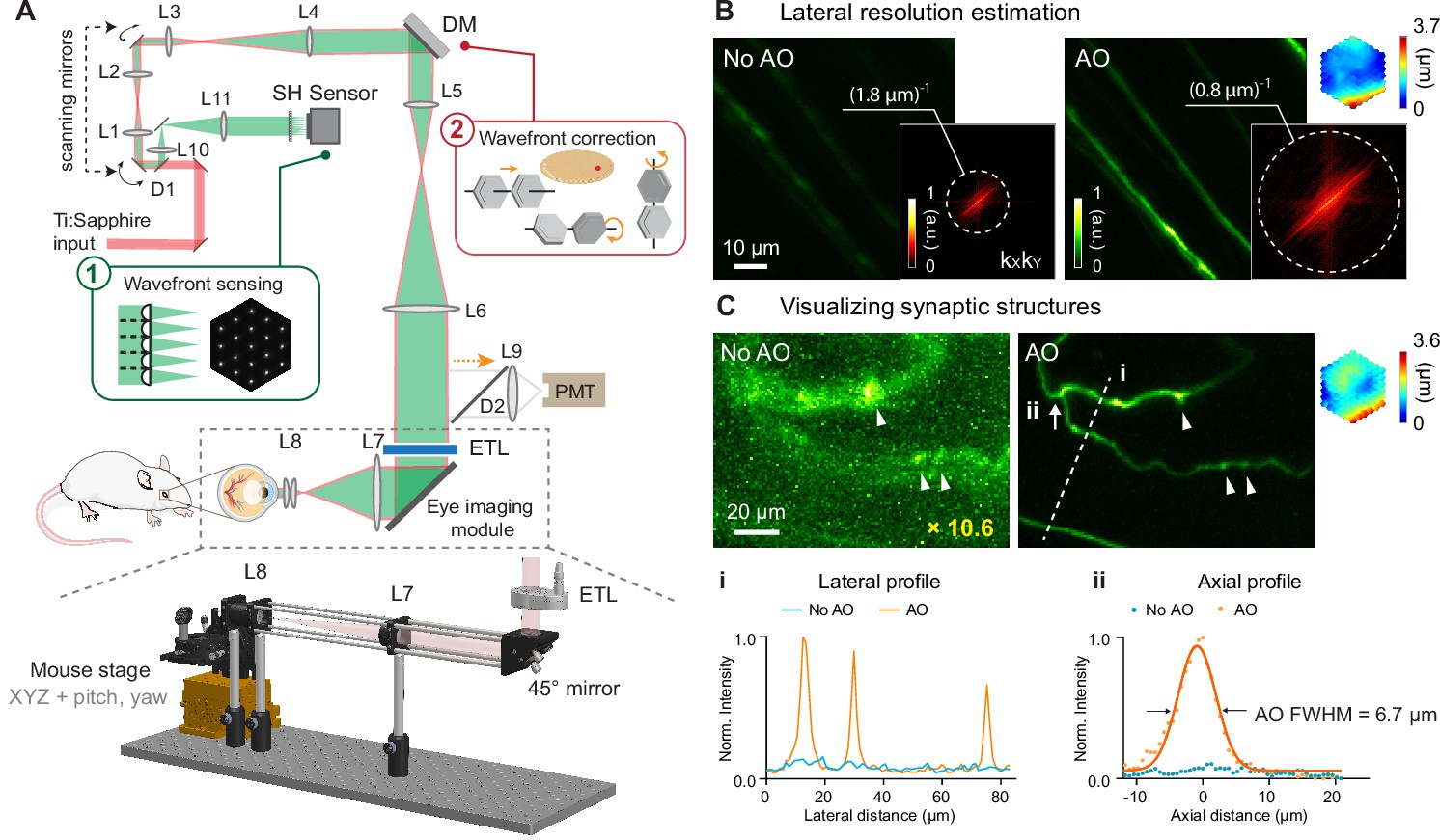
Retinal microvascular and neuronal pathologies probed in vivo by adaptive optical two-photon fluorescence microscopy
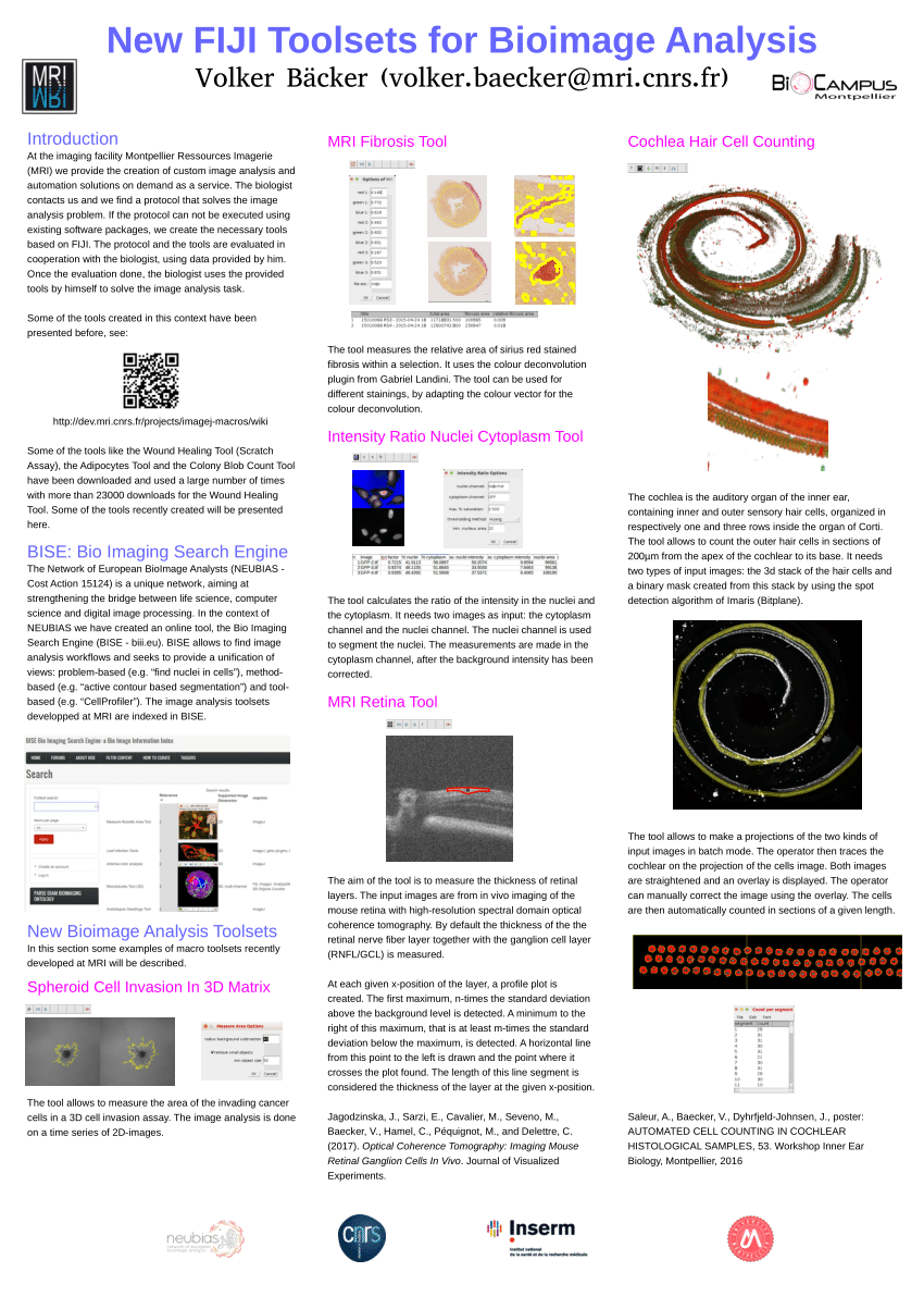
PDF) New FIJI Toolsets for Bioimage Analysis

All Protocols and Video Articles in JoVE

Fig. 9.11, [In vivo confocal reflectance and]. - High Resolution Imaging in Microscopy and Ophthalmology - NCBI Bookshelf
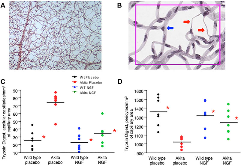
Frontiers Topical nerve growth factor prevents neurodegenerative and vascular stages of diabetic retinopathy

Topical photodynamic therapy combined with ablative “light needles” against basal cell carcinoma - ScienceDirect
Optical Coherence Tomography: Imaging Mouse Retinal Ganglion Cells In Vivo
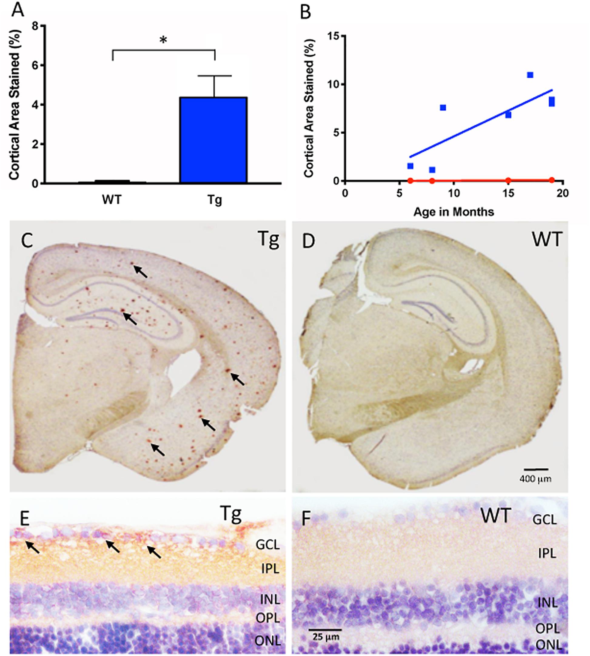
Frontiers In vivo Retinal Fluorescence Imaging With Curcumin in an Alzheimer Mouse Model
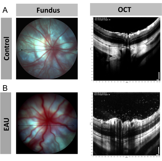
Image-Guided Optical Coherence Tomography to Assess Structural Changes in Rodent Retinas
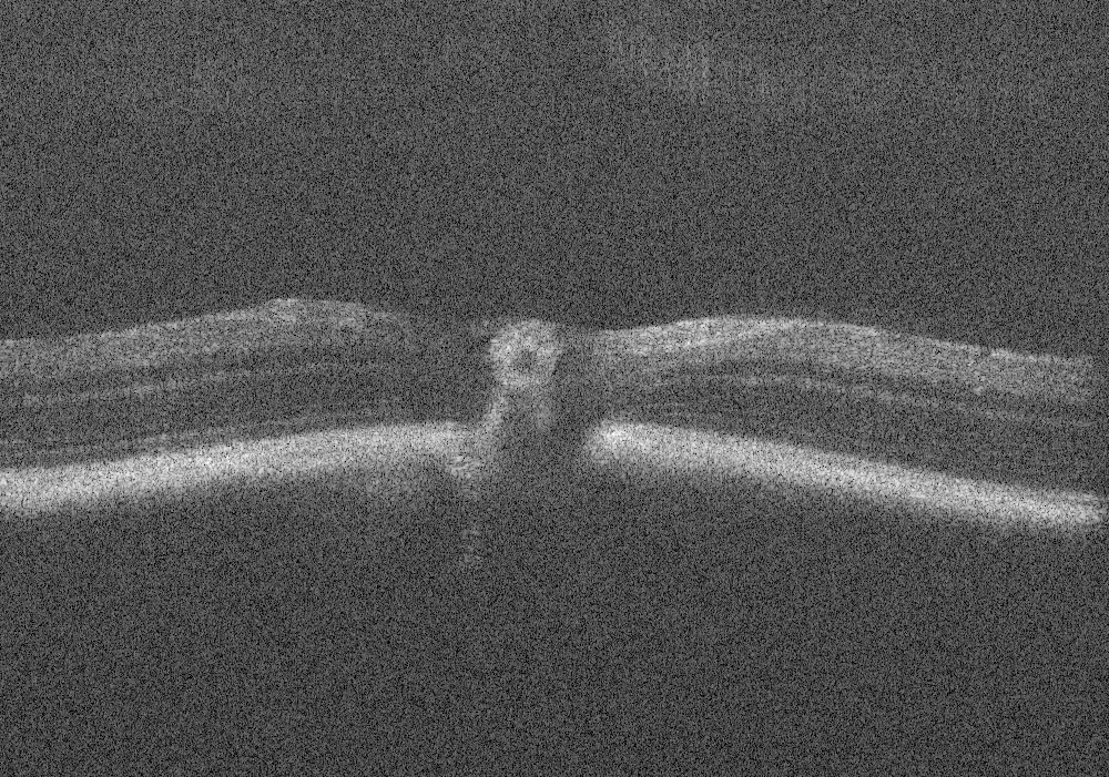
PDF) Early Retinal Defects in Fmr1−/y Mice: Toward a Critical Role of Visual Dys-Sensitivity in the Fragile X Syndrome Phenotype?
Retina Tool - ImageJ-macros - MRI's Redmine

SLO images of the retina of a B6.Thy1-YFP-H mouse in vivo. (a)

PDF] Quantitative Analysis of Mouse Retinal Layers Using Automated Segmentation of Spectral Domain Optical Coherence Tomography Images.
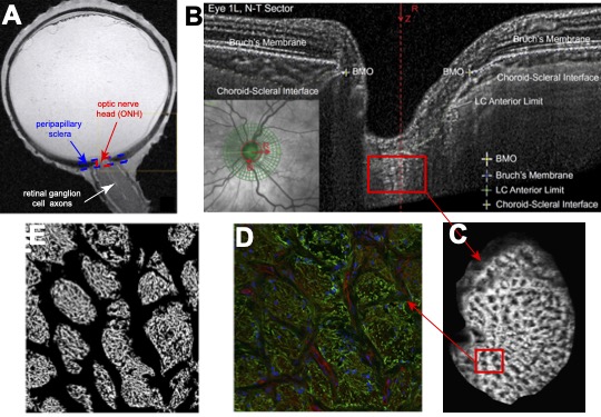
Biomechanics of the Optic Nerve Head – Nguyen Lab
from
per adult (price varies by group size)