Innervation patterns of type I and type II auditory nerve fibers on
By A Mystery Man Writer
Description
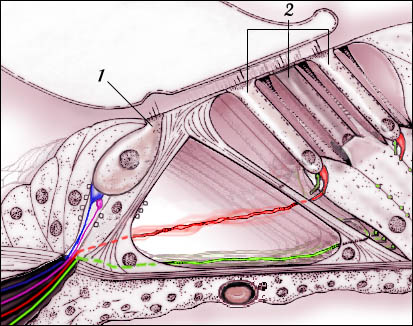
Download scientific diagram | Innervation patterns of type I and type II auditory nerve fibers on inner and outer hair cells, respectively. Central and peripheral axons of type I cells are myelinated, whereas axons of type II neurons are unmyelinated. Peripheral terminals of type I and type II cells are unmyelinated within the organ of Corti, i.e. beyond the habenula perforata. from publication: Noise-induced and age-related hearing loss: New perspectives and potential therapies | The classic view of sensorineural hearing loss has been that the primary damage targets are hair cells and that auditory nerve loss is typically secondary to hair cell degeneration. Recent work has challenged that view. In noise-induced hearing loss, exposures causing only | Hair Cell, Hearing Loss and Neuro-Otology | ResearchGate, the professional network for scientists.
Peripheral Nervous System Anatomy: Overview, Gross Anatomy, Microscopic Anatomy

Cochlear Nucleus

High-resolution volumetric imaging constrains compartmental models to explore synaptic integration and temporal processing by cochlear nucleus globular bushy cells

Profound hearing loss (HL) rates by study. Profound HL rate for

Overview of the processes involved in developing clinical practice
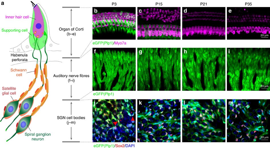
Neuronal heterogeneity and stereotyped connectivity in the auditory afferent system

Electron Microscopic Reconstruction of Neural Circuitry in the Cochlea - ScienceDirect
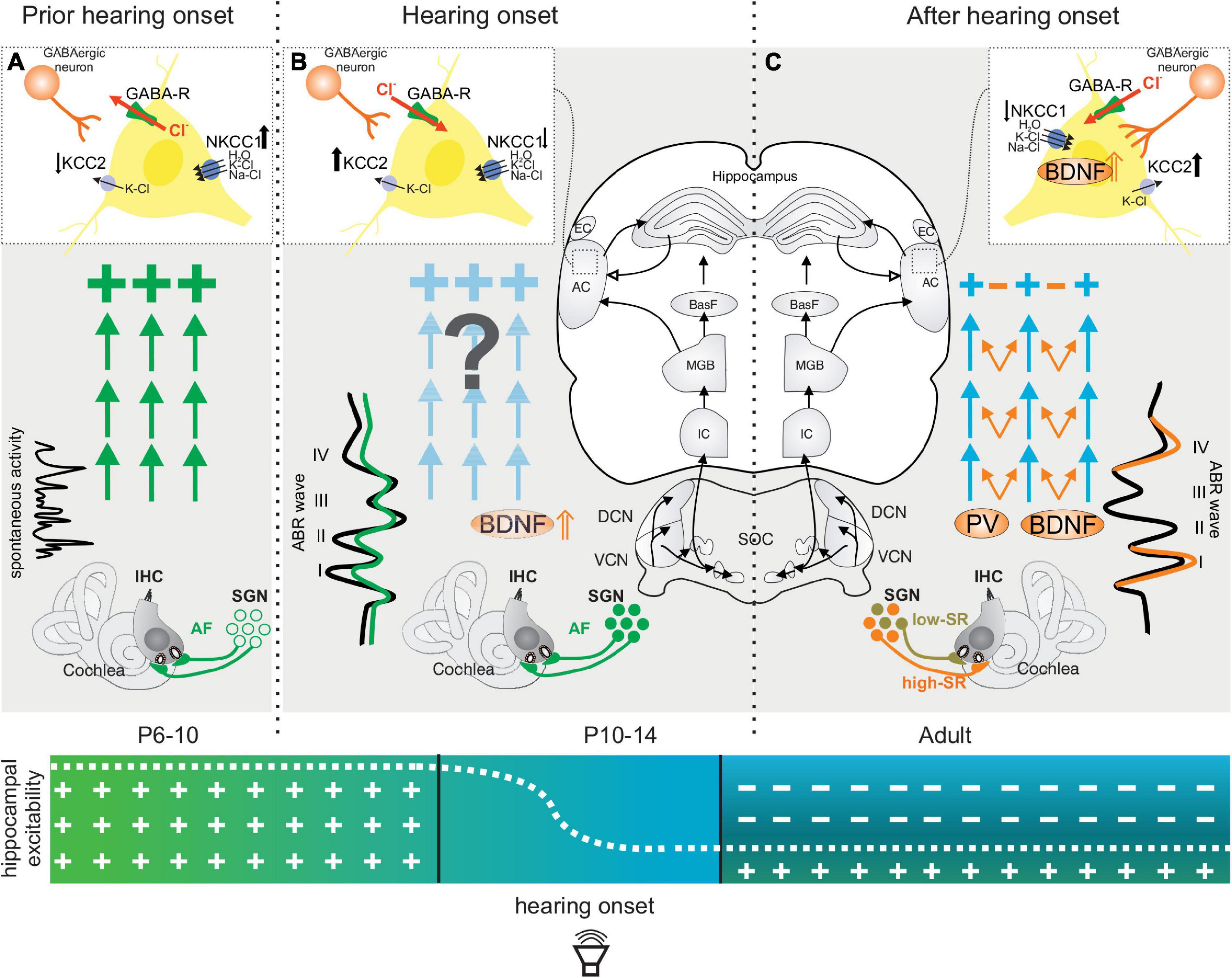
Frontiers Disturbed Balance of Inhibitory Signaling Links
Transient auditory nerve demyelination as a new mechanism for hidden hearing loss

A choreography of intracellular Ca2+ and extracellular ATP to refine auditory nociceptors before hearing
Organ of Corti: innervation
from
per adult (price varies by group size)