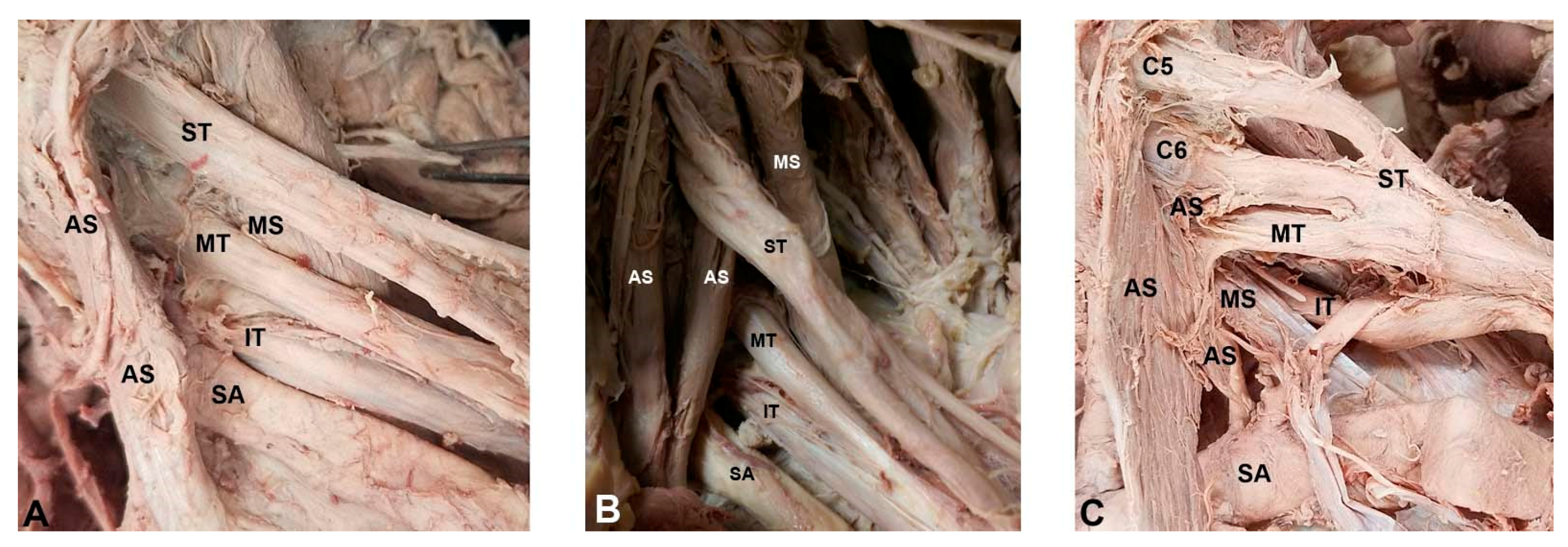
Figure 3 from Descriptive anatomy of the interscalene triangle and
By A Mystery Man Writer
Description
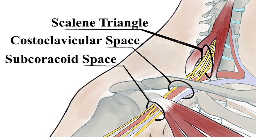
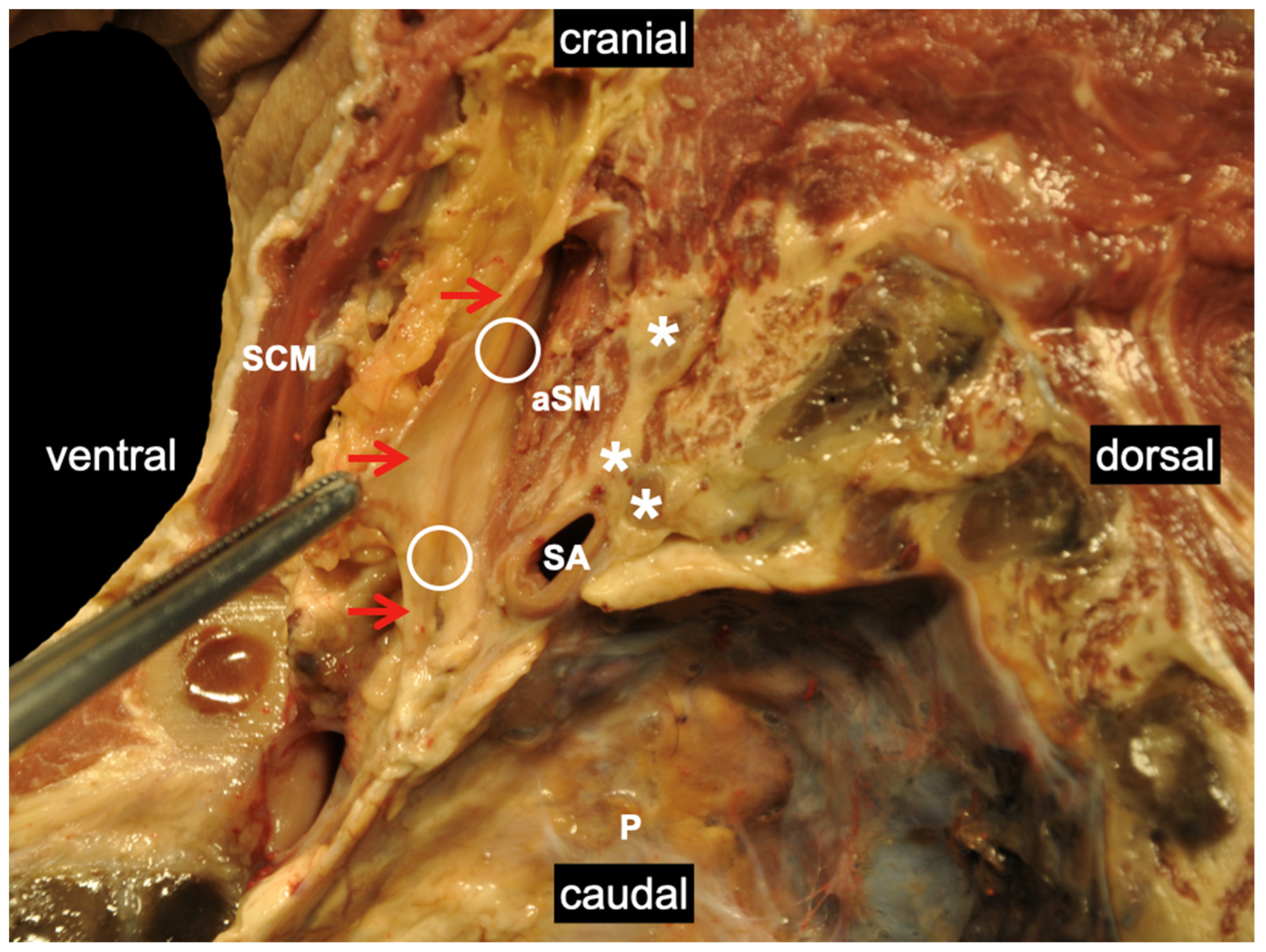
Fig 3. Depiction of the costoclavicular space. The neurovascular elements of the costoclavicular space can be seen here traveling superior to the first rib and inferior to the clavicle. The arrow indicates where measurements were taken. - "Descriptive anatomy of the interscalene triangle and the costoclavicular space and their relationship to thoracic outlet syndrome: a study of 60 cadavers."
Surgical Techniques: Operative Decompression Using the Supraclavicular Approach for Neurogenic Thoracic Outlet Syndrome
Diagnostics, Free Full-Text

Medicina, Free Full-Text

Figure 3 from Descriptive anatomy of the interscalene triangle and the costoclavicular space and their relationship to thoracic outlet syndrome: a study of 60 cadavers.

Diagnostics, Free Full-Text

Modern Treatment of Neurogenic Thoracic Outlet Syndrome: Pathoanatomy, Diagnosis, and Arthroscopic Surgical Technique - ScienceDirect
Historic Basis for the New Developments in the Diagnosis and Treatment of Thoracic Outlet Syndrome (TOS) - Clinical Surgery Journal (ISSN 2767-0023)
Cureus Hydrodissection for the Treatment of Vascular Thoracic

Functional Regional Anesthesia Anatomy.
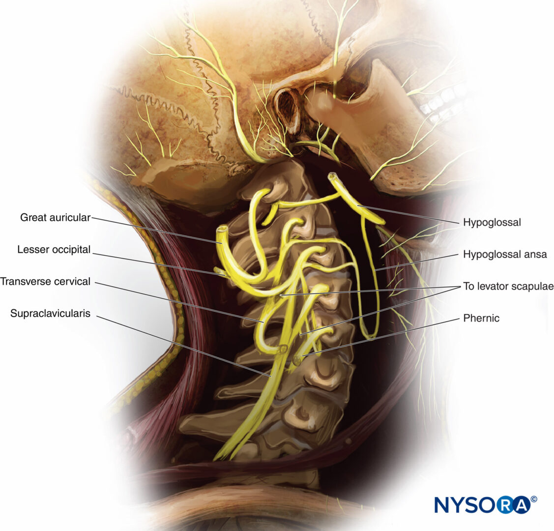
Functional Regional Anesthesia Anatomy - NYSORA

Anatomy, Imaging, and Pathologic Conditions of the Brachial Plexus
Triangles of the neck: Anatomy, borders and contents
Middle Scalene - Physiopedia
Medicina, Free Full-Text
from
per adult (price varies by group size)